Background
Photoelectron spectroscopy was reported by Kai M. Siegbahn in the 1950s. At that time, he reported the photoelectron spectrum of Cu 1s using Mo Kα radiation¹⁾. Subsequently, there were reports of hard X-ray photoelectron spectroscopy in the 1970s²⁾, but due to technical challenges such as insufficient intensity of the hard X-ray excitation source and inadequate photoelectron collection capability of the analyzer, the technique did not reach a practically viable level as an analytical method. As a result, photoelectron spectroscopy was put into practical use using soft X-rays such as Al Kα and Mg Kα radiation, as well as ultraviolet light sources such as helium discharge lamps, and continues to be widely used today as a powerful analytical technique.
With the advent of the 21st century, the emergence of third-generation synchrotron radiation facilities represented by SPring-8 made it possible to utilize hard X-rays with dramatically increased intensity. At the same time, improvements in the voltage tolerance and sensitivity of analyzers resolved the aforementioned technical challenges. As a result, the number of reports on hard X-ray photoelectron spectroscopy³⁾⁴⁾⁵⁾ increased rapidly.
Reports on hard X-ray photoelectron spectroscopy continue to increase to this day, attracting attention as a relatively new analytical technique.

Principles of hard X-ray photoelectron spectroscopy
The basic principle of hard X-ray photoelectron spectroscopy is the same as conventional XPS: exciting the sample surface with excitation light and measuring the kinetic energy of the emitted photoelectrons. While the photon energy of monochromatic Al Kα radiation, most commonly used in conventional
Hard X-ray photoelectron spectroscopy is written as “Hard X-ray Photoelectron Spectroscopy” and is abbreviated as HX-PES or HAXPES. To distinguish it from this, photoelectron spectroscopy using soft X-rays such as Al Kα and Mg Kα radiation as excitation sources is sometimes referred to as “Soft X-ray Photoelectron Spectroscopy” (abbreviated as SX-PES).
Features of hard X-ray photoelectron spectroscopy
Information from deep core levels
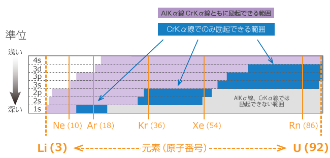
Since hard X-rays have high incident energy, they can excite photoelectrons from deeper core levels than soft X-rays. For example, in the case of Si, soft X-rays can only excite up to the 2s orbital, whereas hard X-rays can excite up to the 1s orbital. Figure 1 shows examples of photoelectron excitation using Al Kα (hν = 1486.6 eV) and Cr Kα (hν = 5414.8 eV) radiation.
Fig 1. Schematic diagram of photoelectron excitation of Si
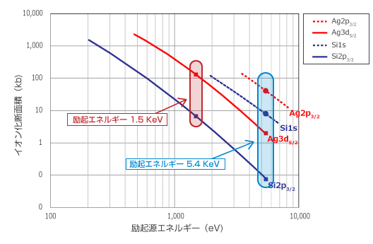
While photoelectron excitation from deep core levels becomes possible, the photoionization cross-section decreases significantly as the excitation energy increases. The photoionization cross-section is a physical quantity related to the process of photoelectron excitation, and the intensity of spectra obtained by photoelectron spectroscopy is proportional to this value. Figure 2 shows the photoionization cross-sections of Si and Ag plotted against excitation energy. For Si 2p3/2 and Ag 3d5/2, which are mainly measured with Al Kα radiation, the photoionization cross-section decreases by one to two orders of magnitude when the excitation energy increases from 1.5 keV to 5.0 keV. However, for Si 1s and Ag 2p3/2, which can be measured with Cr Kα radiation, the decrease in photoionization cross-section with increasing excitation energy is small, and for Si 1s, the ionization cross-section is nearly equivalent at excitation energies of 1.5 keV and 5.0 keV.
In this way, hard X-ray photoelectron spectroscopy offers more measurable core levels than soft X-rays, making it possible to select the optimal level for the purpose, such as avoiding overlapping peaks or selecting deeper core levels.
Information from deeper regions
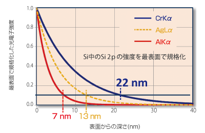
The escape depth of photoelectrons is equivalent to the inelastic mean free path (IMFP) of electrons within a material, and the information depth of photoelectrons obtained as a signal is said to be approximately two to three times the IMFP. A key feature of hard X-ray photoelectron spectroscopy is that the use of higher energy X-rays enables information to be obtained from deeper regions.
For example, when comparing Al Kα and Cr Kα radiation, Cr Kα provides information from depths more than three times deeper than Al Kα. In measurements using Cr Kα radiation, the influence of contamination, adsorbed species, and native oxide layers present within a few nm of the surface is relatively reduced, enabling photoelectron spectra that more closely reflect the true sample information. In addition, buried interfaces within samples can be analyzed non-destructively, and great expectations are held for samples that were difficult to evaluate with Al Kα radiation.
References
- E. Sokolowski, C.Nordling and K.Siegbahn : Arkib för Fysik, 12(1957),
301; Phys.Rev., 105(1957), 1676. - I. Lindau, P. Pianetta, S. Doniach, and W. E. Spicer, Nature,
250, 214 (1974). - T. Ishikawa, K. Tamasaku, and M. Yabashi, Nucl. Instrum. Methods
Phys. Res., A547, 42 (2005). - K. Kobayashi, M. Yabashi, Y. Tanaka, T. Tokushima, S. Shin, K. Tamasaku, D. Miwa, T. Ishikawa, H. Nohira, T. Hattori, Y. Sugita, O. Nakatsuka, A. Sakai, and S. Zaima, Appl. Phys. Lett., 83, 1005 (2003).
- C. Dallera, L. Duò, L. Braicovich, G. Panaccione, G. Paolicelli, B. Cowie, and J. Zegenhagen, Appl. Phys. Lett., 85. 4532 (2004).